- Muscle movement begins at the microscopic level: every contraction results from the precise interaction of nerve signals, chemical reactions and mechanical processes.
- The sliding filament model: actin and myosin filaments slide past one another, shortening the sarcomere – the smallest functional unit of muscle.
- Calcium and ATP control the process: calcium clears the way for myosin, while ATP provides both the energy and the “switch” for releasing and reloading the myosin heads.
- Horse muscles are highly specialised: they feature exceptionally fast calcium dynamics, a high myofibril density and versatile muscle fibres – ideal for precise, powerful movements.
- Efficient energy use and lactate utilisation: horses can actively use lactate as an energy source, which enhances both stamina and performance.
The fascinating world of muscle contraction in horses
The elegant yet powerful movement of a horse begins deep within its muscles, at the microscopic level. Every muscle contraction – whether in the gentle lift of a hoof or the powerful take-off over an obstacle – is the result of a highly complex interplay of nerve signals, chemical reactions and mechanical processes. At the heart of this fascinating process is the sliding filament model – a finely tuned mechanism in which protein structures within the muscle fibre slide against one another, resulting in muscle shortening.
In horses, these processes are organised with exceptional efficiency – the result of thousands of years of evolutionary adaptation as running animals. These physiological specialisations make horses outstanding athletes and set them apart clearly from other mammals.
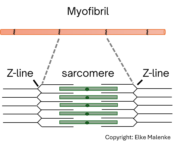
The sarcomere: the smallest unit of muscle contraction
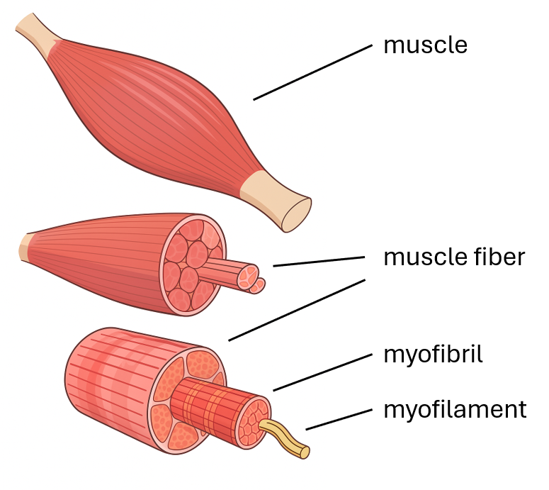
The muscle fibre forms the functional basic unit of the entire musculature and contains hundreds of so-called myofibrils. These are made up of regularly arranged protein filaments: actin (thin filaments) and myosin (thicker filaments with movable heads). Together, these filaments form the sarcomere – the smallest contractile unit of muscle.
Thousands of these sarcomeres are linked together like beads on a string and work in synchrony to produce the visible shortening of a muscle fibre – and thus the overall muscle movement. In the resting state, actin and myosin lie close to one another but are not yet connected. Only an external stimulus in the form of a nerve impulse sets the complex molecular machinery into motion.
From nerve signal to movement: the full process of muscle contraction
Signal transmission and calcium release
Muscle contraction begins with an electrical signal that travels along a motor nerve fibre to the so-called motor end plate of the muscle fibre. There, the important neurotransmitter acetylcholine is released, triggering an electrical excitation at the cell membrane of the muscle fibre. This excitation spreads at lightning speed through specialised membrane structures, the T-tubules, deep into the cell’s interior and activates the sarcoplasmic reticulum – a highly specialised calcium store within the muscle fibre.
The key: calcium and the regulatory proteins
As soon as calcium is released into the cell fluid, it binds to the regulatory protein troponin, which is closely linked to tropomyosin. In the resting state, tropomyosin acts like a protective shield, blocking the binding sites on the actin filament for the myosin heads and thus preventing unwanted contraction. When calcium binds to troponin, this causes tropomyosin to shift position, uncovering the binding sites on actin so that myosin can attach, and contraction can begin.
The contraction itself: the power stroke
Now begins the remarkable process of actual muscle contraction. The myosin heads, already loaded with ATP, attach to the now accessible actin. The release of a phosphate molecule frees chemical energy, causing the myosin head to perform a powerful movement that pulls the actin filament a little further along. This action, known as the power stroke, results in a minimal shortening of the sarcomere.
This fascinating process is repeated in rapid succession, sarcomere by sarcomere, ultimately leading to the visible shortening of the entire muscle. To detach from the actin after the power stroke, the myosin head once again requires ATP. Only with this universal energy molecule can it release, return to its original position, and prepare for the next power stroke.
Relaxation: the return to the resting state
Once the nerve impulse ends, calcium is actively pumped back into the sarcoplasmic reticulum – a process that also requires energy. Tropomyosin once again blocks the binding sites on actin, and the muscle fibre returns to its relaxed state. If ATP is absent – for example, after death – the myosin remains permanently bound to actin. This is the biochemical basis of what is known as rigor mortis.
ATP: the essential fuel for muscle contraction
ATP (adenosine triphosphate) is the body’s central energy transfer system and plays an especially critical role in muscle function. It is required not only for the powerful contraction of the muscle filaments, but also for the release and “reloading” of the myosin heads, as well as for the active return of calcium to its stores.
The amount of immediately available ATP in a muscle cell is very limited and sufficient for only a few seconds of maximal contraction. To enable continuous muscle work, ATP must therefore be constantly regenerated. Depending on the duration and intensity of the workload, this happens via different metabolic pathways: very short, high-intensity efforts draw on the creatine phosphate system for instant ATP supply; medium-intensity efforts rely on anaerobic glycolysis; while longer, endurance activity depends on the aerobic oxidative breakdown of glucose, lactate or fatty acids.
The unique aspects of muscle contraction in horses
Muscle contraction in horses follows the same basic biochemical mechanisms as in other mammals, but it also shows some remarkable specialisations that explain the exceptional performance and resilience of equine musculature.
Ultra-fast calcium control for precise movements
A key feature of the horse musculature is the rapid and efficient calcium dynamics within the muscle fibres. Horses possess particularly powerful sarcoplasmic reticula, which can release calcium extremely quickly and just as rapidly take it back up. This swift alternation between contraction and relaxation allows finely tuned movement patterns and contributes to the high stride frequency seen especially in canter or in complex lateral movements.
Maximum strength from a high density of myofibrils
The high density of myofibrils in horse muscle fibres is exceptional and clearly surpasses that of many other mammals. This unusually high packing density of contractile elements means that each muscle fibre can generate significantly more force. The result is muscles that are not only strong but also extremely efficient – a crucial requirement for carrying a rider’s weight while maintaining full functional capacity.
Type IIa fibres: the best of both worlds
Another distinctive feature lies in the distribution and function of muscle fibre types. Type IIa fibres, which are particularly common in horses, combine strong aerobic endurance with the ability to contract quickly. This unique combination makes them ideal for medium-length, high-intensity efforts, such as those seen in training, competitions or cross-country work. Horses can activate these fibres over extended periods without shifting entirely into anaerobic work, which reduces fatigue and extends performance capacity.
Unique lactate management
In the field of energy metabolism, equine musculature shows further specific adaptations. Its capacity for lactate utilisation is particularly pronounced: horses can not only buffer the lactate produced during intense exertion but also actively use it as an additional energy source. This process, known as “lactate shuttling”, allows lactate to be used as fuel, for example in the heart muscle or in slow-twitch fibres. This ability significantly enhances endurance performance and reduces the risk of lactic acid build-up in the muscles.
Training adaptations: how horse muscles build
A horse’s musculature shows an impressive ability to adapt through targeted training. It responds in highly specific ways to different types of workload – structurally, by increasing muscle cross-sectional area, and functionally, through changes in fibre profile or enzyme activity. With systematic, well-designed training, it is therefore possible not only to improve strength and stamina, but also to optimise movement coordination, muscle metabolism and recovery capacity.
Breed-specific differences and their practical relevance
Muscle contraction and the associated performance characteristics are strongly influenced by breed, use and management. Thoroughbreds, for example, have a completely different muscle profile from baroque horses or draft horses, which directly affects reaction speed, carrying capacity and stamina. These fundamental differences must be taken into account not only in training plans but also in feeding, recovery and health care.
The takeaway: understanding and using nature’s perfection
Understanding muscle contraction in horses reveals a fascinating interplay of biochemistry, physics and evolutionary adaptation. These complex processes, occurring millions of times per second in every muscle fibre, form the basis of the horse’s impressive athletic performance. Only by understanding these fundamental mechanisms is it possible to shape training, feeding and care in a way that fully supports and preserves the horse’s natural abilities.